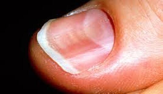
 Koilonychia Nail art and manicure are more popular than before but we don’t give the same importance to nail health. A general examination includes checking the heart rate, BP, blood sugar levels and many more but have we ever taken care to inspect our toenails and fingernails as part of a physical examination? Our nails mirror our internal health and when a physician masters the art of diagnosing nail changes, he/she has the capability to present himself/herself as an excellent diagnostician. The basic requirement for treating a problem is excellent diagnosis and once this is done the problem is as good as almost solved! This was first initiated by Hippocrates when he came up with clubbing by trousseau followed by numerous other systemic associations by many others. Nail Anatomy Each nail includes a nail matrix, the lunula (half-moon shaped area), the nail fold, the nail plate and the nail bed. The nail matrix extends up to the lunula and is responsible for nail plate production. The nail bed extends from the lunula up to the free edge of the nail and gets its reddish colour from optimal blood supply. Most changes or problems that can be diagnosed include changes to the nail plate (discoloration, lines, bands, grooves, altered shapes and attachment to nail bed), nail bed or nail folds. While systemic diseases impact nail matrix growth diabetes, drugs and sepsis affect nail folds. Genetic disorders too affect nail growth but the changes in nail anatomy caused due to internal changes is more difficult to diagnose and needs keen observation. All the 20 nails are clearly visible to us and this clarity makes it easier to observe and use them as a diagnostic tool. All the nails must be observed after removal of nail polish (if present) to enable correct diagnosis. Nail Shape Abnormalities Koilonychia: Also known as spoon-shaped nails (as the nail shape is so bad that it can contain a drop of water), this is a reverse curvature in the transverse and longitudinal axis of nail plates giving them a concave appearance. This might be due to conditions such as iron-deficiency anaemia (commonly seen), malnutrition, thyroid, coronary disease, traumatic injury, upper gastrointestinal malignancy or occupational (commonly seen). Mostly seen in infants it disappears during growing years but old age and digital ischemia are also common causes for the disease. Clubbing: There is an increased nail plate curvature longitudinally and transversely with soft tissue hypertrophy of the digital pulp involving all 20 digits. As this problem is associated with innumerable conditions it’s been split into three major categories: idiopathic, hereditary-congenital and acquired. Systemic diseases such as cyanotic congenital heart disease, lung cancer, infective endocarditis, lung abscess, inflammatory bowel disease, cystic fibrosis and hepatic cirrhosis exist as risk factors. It might also be signs of AIDS in HIV-positive kids. Dolichonychia: Here, the length of nails surpasses the width of nail. This condition is mostly associated with Marfan’s syndrome and hypopituitarism. Brachyonychia: Here, the width of the nail is small compared to the length being a sign of hyperparathyroidism and psoriatic arthropathy indicating early bone resorption. Parrot beak nail: The edge of the fingernails are deformed in the shape of a parrot’s beak (hence the name) bending around the shortened fingertip. It is seen in severe acrosclerosis with distal phalangeal resorption due to scleroderma. Macronychia & Micronychia: The nails of the affected finger are either too large or too small compared to other nails nearby. While Macronychia might be due to local gigantism Micronychia may be linked to plexiform neuromas.
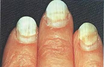
Problems with Nail Attachments
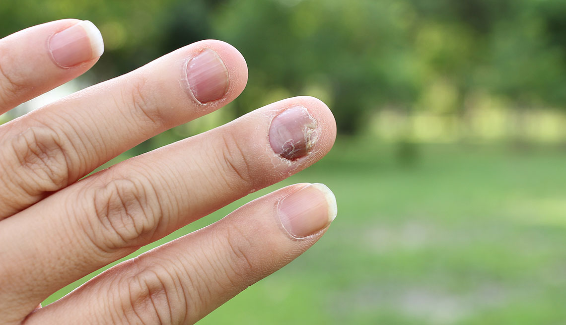
Onycholysis: The nail plates and nail beds are separated from each other and the areas of separation appear yellow or white due to presence of air beneath the nail and sequestered debris. The cause of origin is more alongside conditions such as trauma, psoriasis, fungus and contact irritant reaction than systemic diseases such as thyroid, vitamin C deficiency, psoriatic arthritis, lupus, lung cancer, T-cell leukaemia and anaemia. Pterygium: This involves scarring of the nail matrix classically seen in lichen planus and commonly seen in burns, graft versus host disease, radiodermatitis, lupus and cicatricial pemphigoid. Conditions Associated with Nail Surface Longitudinal Ridging: Do you see vertical lines occupying your entire nail surface? Then these imply long-lasting abnormalities such as nail-patella syndrome, systemic amyloidosis, collagen vascular diseases, graft versus host disease and rheumatoid arthritis or it might also be due to a minor trauma (this is the root cause in most cases). Central ridges might also be due to iron, folic acid or protein deficiency. Beau’s lines: These are not true lines but transverse grooves in the nail plates arising from temporary suppression of nail growth within the nail matrix when an individual is under stress or faced with systemic illnesses such as heart attack, fever, exposure to extreme cold, stress and poor nutritional status. If in case the grooves are present in all 20 nails it is the result of systemic disease such as mumps, pneumonia, coronary thrombosis, Kawasaki disease, syphilis and hypoparathyroidism. Nail pitting: Here the keratin layer is not intact in the proximal matrix with a number of parakeratotic cells in the nail plate surface. Onychochizia: Trauma leads to horizontal splitting of the nail toward its distal portion called as lamellar splitting of nails. Changes in Nail Colour Leukonychia: if you notice white discoloration of nails it must be leukonychia which can be subdivided into three types namely true, apparent and pseudo. It is either true or apparent that is mostly linked to systemic diseases. Mees lines: These are present as traverse white lines 1-2 mm wide horizontally spanning the width of the nail plate and occurring in all fingernails. Mostly due to arsenic poisoning it helps the patient uncover the exact time of the poisoning as the lines appear two months after the incident. Other causes include renal failures, breast cancer, ulcerative colitis, infections such as measles and tuberculosis, lupus and exposure to toxic metals such as thallium. Muehrcke lines: These are double white transverse lines covering the width of the nail bed running parallel to the distal lunula. These lines are mostly reported in patients suffering from liver disease, malnutrition, chemotherapy, organ transplant, HIV and acquired immunodeficiency syndrome. They might also be due to metabolic stress when the patient lacks the ability to synthesize proteins. Lindsay nail: This is leukonychia with a normal proximal half and abnormal brownish discoloured distal half commonly seen in patients with chronic kidney disease having uremic renal failure. Terry nail: Leukonychia having the presence of white proximal and normal distal nails associated with congestive cardiac failure, adult-onset diabetes mellitus, peripheral vascular disease, hemodialysis and HIV. Melanonychia: This is the appearance of brown-black vertical lines in the nail plate due to trauma, bacterial, fungal or HIV infection, drug therapy, endocrine disorders, exogenous pigmentation and excess melanin production. Cyanosis: A blue or purple discoloration of the nail bed due to decreased oxygen supply causes accumulation of deoxyhaemoglobin in the small blood vessels of the extremities. This might occur as a result of cold, congestive cardiac failure and peripheral vascular disease. Icterus: This manifests in the form of yellow discoloration of the mucosae due to bilirubin deposition in extreme cases representing a severe form of liver disease. Splinter haemorrhages: This is due to extravasation of blood from the longitudinal blood vessels of the nail bed due to psoriasis and in some cases due to infective endocarditis and rheumatic heart disease. Yellow nail syndrome: Mostly linked to systemic diseases such as lymphedema and compromised respiration (due to pleural effusion) it leads to yellow or yellow-green discoloration of the nails. Red lunula: Red coloration might be visible in the distal region of the lunula in the nail bed or be demarcated by a pale line commonly occurring as a result of collagen vascular disease, cardiac failure, chronic obstructive pulmonary disease (COPD), cirrhosis, psoriasis and CO poisoning. Inherited Diseases: Sometimes, rare diseases such as Hailey-Hailey disease (mutation of ATP2A2 gene) and Darier disease (mutation of ATP2C1 gene) lead to white longitudinal bands in the nails that’s of varied widths besides having vesicular eruptions in the flexural skin area including the neck, groin and axilla. Some patients (92-95% patients) with Darier disease might have alternating streaks of red and white called as ‘candy cane’. Glomus Tumour: This is a benign neoplasm that originates from a neuromyoarterial glomus body (located all through the body but available in greater numbers at the fingertips mostly beneath the nails). This makes nails a viable place for glomus tumor. The nails suffer from intense, pulsatile pain that’s sensitive to cold and there is tenderness after pin-point palpation of the tumor. Onychopapilloma: This is a benign idiopathic tumor that prevails as the common cause behind localized longitudinal erythronychia. Asymptomatic, less common benign conditions include warts, benign vascular proliferation, a solitary lesion of lichen planus and postsurgical scarring of the nail matrix. Polydactyl erythronychia occurs as red streaks in multiple nails and exists as the main symptom behind lichen planus or Darier disease. Sometimes it is also linked to systemic amyloidosis, hemiplegia, graft-vs-host disease or pseudobulbar syndrome. References Nail as a Window of Systemic Diseases: https://www.ncbi.nlm.nih.gov/pmc/articles/PMC4375768/ Evaluation of Nail Lines: Colour & Shape Hold Clues: https://mdedge-files-live.s3.us-east-2.amazonaws.com/files/s3fs-public/issues/articles/Lipner_NailLines.pdf Nail Evaluation in Internal Diseases: An Indispensable Exercise: http://www.amhsjournal.org/article.asp?issn=2321-4848;year=2017;volume=5;issue=2;spage=269;epage=274;aulast=Gopal
0 Comments
Leave a Reply. |
AVOID FRAUD. EAT SMART.
+91 7846 800 800
AuthorDietitian & Nutritionist Dr. Nafeesa Imteyaz. Archives
July 2024
Categories
All
Dr. Nafeesa's Blog @blogspot |
- Home
- Written Testimonials
- Consult
- Clinics
- Blogs
-
Diet & Nutrition
- Diabetes Reversal
- IVF IUI not needed for PCOS PCOD Infertility
-
Medical Nutrition
>
-
Disease & Conditions
>
- Infertility | PCOS
- Diabetes Mellitus
- Cholesterol
- Hypothyroid
- Kidney Problems
- Hypertension
- Cardiovascular Diseases
- Liver Diseases
- Gastro intestinal disorder
- Cancer
- Metabolic Disorders
- Orthopedic Disorders
- Eating Disorders
- Dietary Recall
- Weight Record Filled By Clients
- Online Payment Transaction Details
- Online Clients Weight Check Form
- Our Program Package Service Charges
- Weight Record 2017 Clients
- Measurements sent by Clients
- Terms & Conditions Of Payment
- Thanks. Your Form is Submitted
- Video Testimonials
- Lifestyle & Wellness
- Lifestyle & Wellness Blog
- Allergy & Intolerance
- Weight Loss / Gain
- Weight Loss / Slimming Blog
-
Disease & Conditions
>
- Life Cycle Nutrition >
- Sports Nutrition >
- Integrity in Nutrition
- Knowledge Centre
© COPYRIGHT 2022. ALL RIGHTS RESERVED. FRST HEALTHCARE PVT LTD.
Dr. Nafeesa Imteyaz of First Eat Right clinic, is the Best Dietitian Nutritionist in Bangalore. Best Dietitian Nutritionist in Pune. Best Dietitian Nutritionist in Hyderabad. Best Dietitian Nutritionist in Chennai. Best Dietitian Nutritionist in Mumbai. Best Dietitian Nutritionist in Delhi. Best Dietitian Nutritionist in Kolkata.